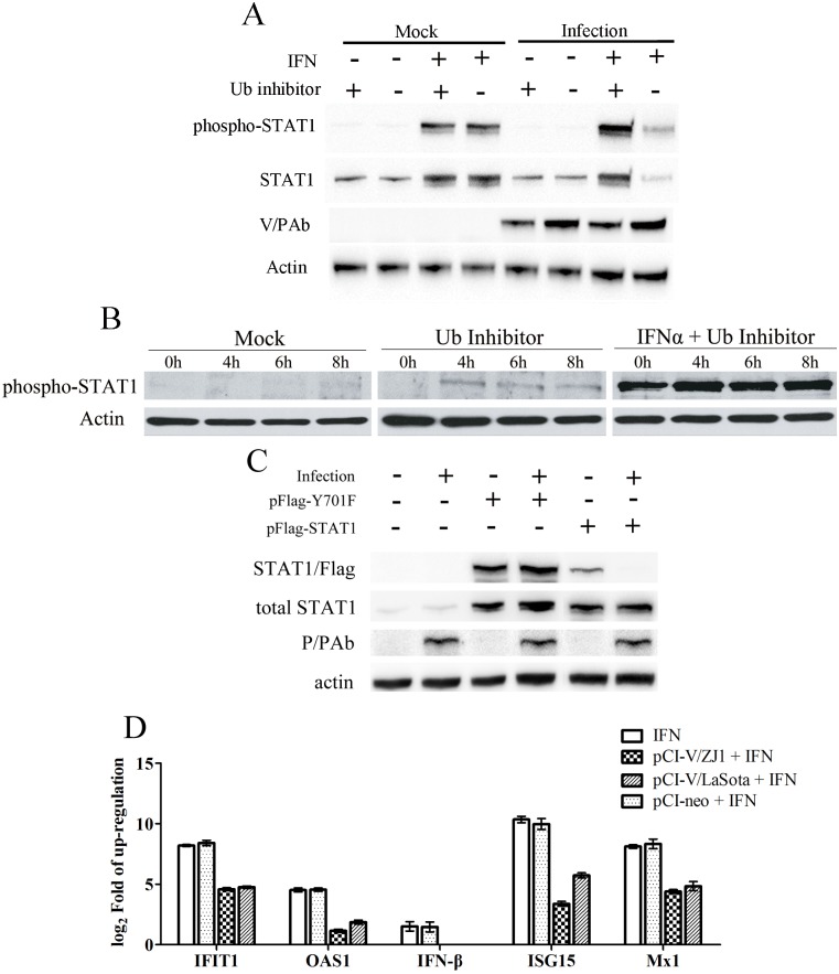
Fig 3. The reduction of total and phosphorylated STAT1 in NDV-infected cells was inhibited after treatment with Ub E1 inhibitor PYR-41 at different time points.
(A) STAT1 and phospho-STAT1 expression levels in PYR-41 treated Vero cells at 6 hpi. (B) Phospho-STAT1 levels in PYR-41 treated Vero cells at 4, 6, 8 hpi. (C) Exogenous mutant STAT1 lacking 701aa phosphorylation site were not degraded in the course of NDV infection. One microgram pFlag-STAT1 or pFlag-Y701F was transfected into A549 cells cultured in 6-well plates. At 12 h post transfection, the cells were subsequently infected with NDVs at a MOI of 3. The cells were harvested at 24 hpi. (D) The expression levels of IFN-responsive genes in V-expressing A549 cells after stimulation of IFN-α. A549 cells in 6-well plates were transfected with 3 μg pCI-V or pCI-neo for each well as above-described. At 4 h and 8 h post-transfection, the cells were harvested following the treatment with 500 U/ml IFN-α for 30 min.

