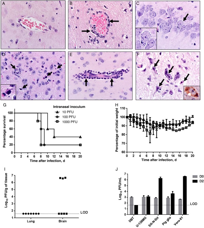
Figure 4.
Brain disease in Middle East respiratory syndrome coronavirus (MERS-CoV)–infected human cytokeratin 18 (K18)–hDPP4 and uninfected mice. A, Normal brain from an uninfected mouse. B, MERS-CoV caused lymphocytic perivascular cuffing in the infected brain. C, Infected neuron in hippocampus 6 days after infection. Note the granular degeneration and basophilic cytoplasmic inclusions (arrow and inset). D, Dying cells undergoing degeneration (arrows and inset; 6 days after infection) are detected in highly infected regions such as the thalamus or brain stem. E, Meningeal and perivascular cuffing included neutrophilic infiltrates (arrows; 6 days after infection). F, Several degenerating cells had small to granular basophilic cytoplasmic inclusions (arrows; 6 days after infection) that were stained with anti–MERS-CoV antibody (inset; brown). Note the neuropil rarefaction. Section were stained with hematoxylin-eosin (original magnification ×600). G–I, Outcomes of K18-hDPP4 mice infected with different intranasal inocula of MERS-CoV. K18-hDPP4 mice received 1000, 100, or 10 plaque-forming units (PFU) of MERS-CoV and were monitored for survival (G) and weight (H). There were 5 mice/group. I, Lungs and brains of mice receiving 10 PFU were harvested 10 days after inoculation or when they lost 20% of body weight. A total of 3 of 7 MERS-CoV–infected mice showed high virus titers in the brains. J, MERS-CoV replicates in cells of the nervous system. Human central nervous system–derived cell lines (U-138 MG an SK-N-SH), primary porcine astrocytes, a murine astrocytoma cell line (DBT), and African green monkey kidney cells (Vero-81) were infected with MERS-CoV at a multiplicity of infection of 1. Titers from these cells immediately after infection (day 0) or 2 days after infection were determined by plaque assay. Data are mean ± SD for 3 replicates/condition. Abbreviation: LOD, limit of detection.

