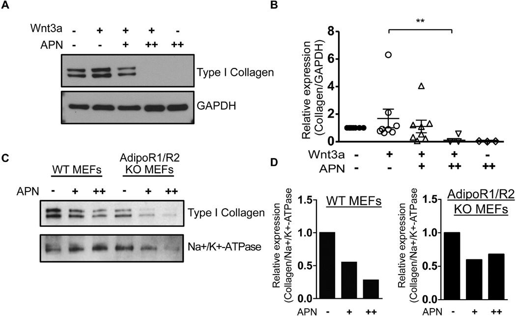
Figure 4. Adiponectin represses Type I Collagen expression independently of AdipoR1/R2 receptors.
A and B) Primary human skin fibroblasts were serum-starved overnight and then treated with adiponectin (APN; 1 or 5 µg/ml). Twenty-four hours later, cells were treated with Wnt3a (100ng/ml). Five days after Wnt3a treatment, lysates were subjected to immunoblot analysis to measure type I collagen. The graph in B shows relative collagen expression from at least four independent experiments. The Mann-Whitney U test shows that Wnt3a causes a statistically significant decrease in collagen (p = .0016). Panel A depicts a representative blot from those graphed in panel B. GAPDH served as a loading control. (C and D) Wild-type (WT) or AdipoR1/R2 KO MEFs were serum-starved overnight and then treated with adiponectin (APN; 5 or 10 µg/ml). Five days later lysates were subjected to immunoblot analysis for type I collagen. Bands shown in panel C are quantified in panel D. Na+/k+-ATPase immunoblot served as a loading control.

