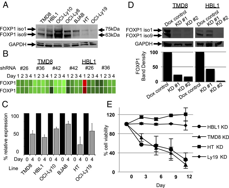
Fig. 1.
FOXP1 is highly expressed in ABC-DLBCL cell lines, and its loss results in their death by apoptosis. (A) Anti-FOXP1 Western blot of whole cell lysates prepared from DLBCL cell lines shows stronger expression of FOXP1 isoforms 1 and 9 in ABC- than in GCB-DLBCL lines. (B) Heat map of FOXP1 probes after FOXP1 knockdown in HBL1 and TMD8 with indicated shRNAs and at indicated time points (microarray accession no. GSE64586). (C) Assessment of FOXP1 knockdown by RT-qPCR normalized to GAPDH in the indicated cell lines after 4 d of doxycycline (dox) (1–10 μg/mL) induction of shRNA. Data include a minimum of four biological repetitions; error bars are SE of the mean. (D) Western blot of FOXP1 KD after 4 d of dox induction in TMD8 and HBL1 cell lines confirms reduction in FOXP1 protein level with two different shRNAs; bar graphs are normalized to GAPDH and compared with nonspecific shRNA control (dox control). (E) Loss of cell viability of ABC- but not GCB-DLBCL lines after 12 d of inducible FOXP1 KD as assessed by constitutive GFP levels independently encoded by the FOXP1 shRNA vector pRSMX.

