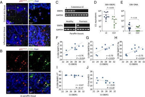
Fig. 7.
EBER1 is present in pDC-infiltrated SLE skin lesion in the absence of EBV-DNA. (A) Immunofluorescence staining of paraffin-embedded inflamed and healthy skin tissue. CD123 is used as pDC marker and CD1a as Langerhans cell (LC) marker. Nuclei are stained with DAPI. (B) Immunofluorescence staining of cutaneous LE paraffin tissues shows CD123+/CD40+ cells. (C) qPCR of EBER1 in lupus erythematosus and control (healthy and psoriasis) tissues. (D and E) qPCR analysis of EBER1 (D) and EBV-DNA (E) in control and cutaneous LE frozen tissues. qPCR data are expressed as EBV+ LCL equivalents. (F–J) Correlation of EBV-miRNAs and hsa-miR-21 with EBER1 in skin tissues analyzed by qPCR.

