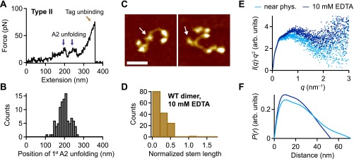
Fig. S9.
Flexibility of VWF dimers induced by addition of EDTA. (A) Denoised force–extension traces of EDTA-treated dimers showing A2 unfolding peaks only at high extension values (type II). The characteristic high-force peak, which was observed for dimers under near-physiological conditions, was never observed. (B) Unimodal distribution of the position of the first A2 unfolding event. (C) Representative AFM images of individual VWF dimers adsorbed from EDTA buffer at pH 7.4. Arrows mark the positions of the CK domains. (Scale bar, 30 ; range of color scale, 2.4 .) (D) Distribution of the normalized stem length of dimers adsorbed from EDTA buffer, showing a single peak decaying from zero stem length. (E) SAXS profiles in Kratky representation [ vs. q] for dimeric A1-CK constructs at pH 7.4 in the presence of divalent ions (light blue) and upon addition of EDTA (dark blue). (F) Distance distribution functions of dimeric A1-CK constructs computed from experimental SAXS data represented in the same colors as in E. The functions are normalized to give equal areas.

