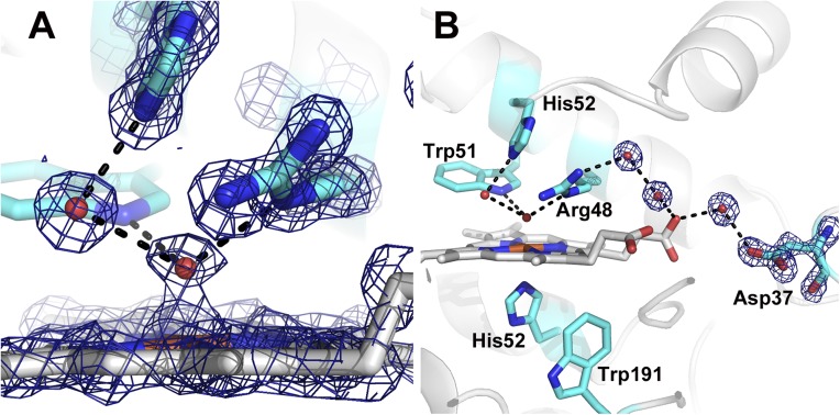
Fig. 2.
2Fo-Fc electron density map of the XFEL CmpI structure contoured at 2.0 σ. The dashed lines indicate H-bonding interactions, which are all less than 3.0 Å. The Fe(IV)-O bond length is 1.7 Å. Close-up view of the ferryl center is shown (A), as well as the extensive H-bonded network connecting the ferryl O atom to the surface of the enzyme (B).

