
Figure 3.
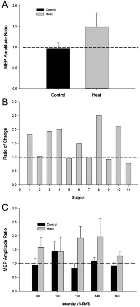
The MEP responses to heat stress. (A) The Ratio (post/pre) of the MEP amplitude measured after 30 minutes of no heat stress (Control) or after 30 minutes of heat stress (Heat) (p < 0.05). (B) The Ratio of the change in MEP amplitude ((Heat (post/pre)/Control (post/pre)) for each subject in the study. Six of the eleven subjects (5 male; subjects 1, 3, 4, 8, 10) showed 49% or greater increase in MEP amplitude ratio after the heat condition compared to the control condition. (C) The MEP amplitude ratio for each intensity assessed during the TMS protocol. There was an overall significant main effect for heat stress in the Heat condition, (P = 0.05).