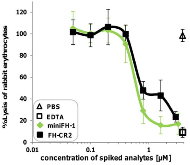
Figure 3. Protection of rabbit erythrocytes from complement AP mediated lysis.
Rabbit erythrocytes were incubated for 30 min in serum (from healthy donors) spiked with complement inhibitors (75% final serum conc.). The lysis of rabbit erythrocytes was measured by hemoglobin release and normalized to lysis observed in water (Average of 3 or 2 independent assays with SD is shown for miniFH-1 and FH-CR2, respectively).

