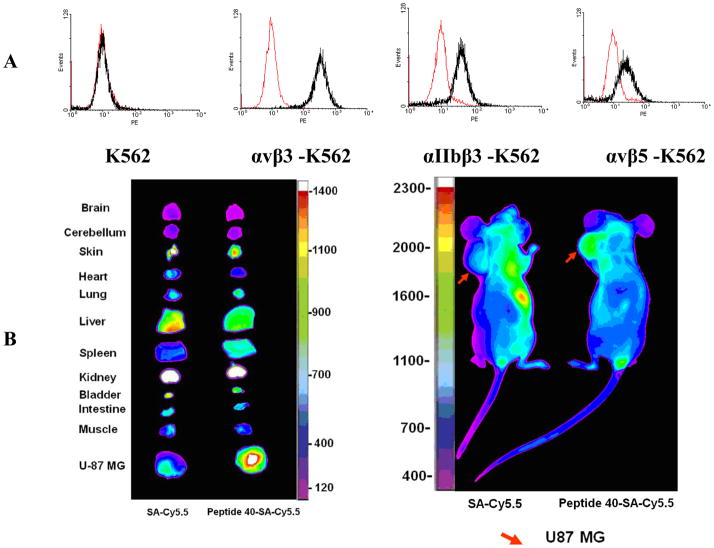
Figure 4.
A. Series of integrin transfected K562 cells were stained with LXW64. Black curves: cells were treated with 1μM biotinylated LXW64 and Streptavidin-PE successively, and measured with Flow Cytometry. Red curves represent cells without treatment of biotinylated LXW64, as negative controls. LXW64 showed significant positive binding with αvβ3, weak cross-reaction with αvβ5 and αIIbβ3, and no binding with α5β1expressed on K562 cells. B. In Vivo and Ex Vivo near-infrared fluorescence images.

