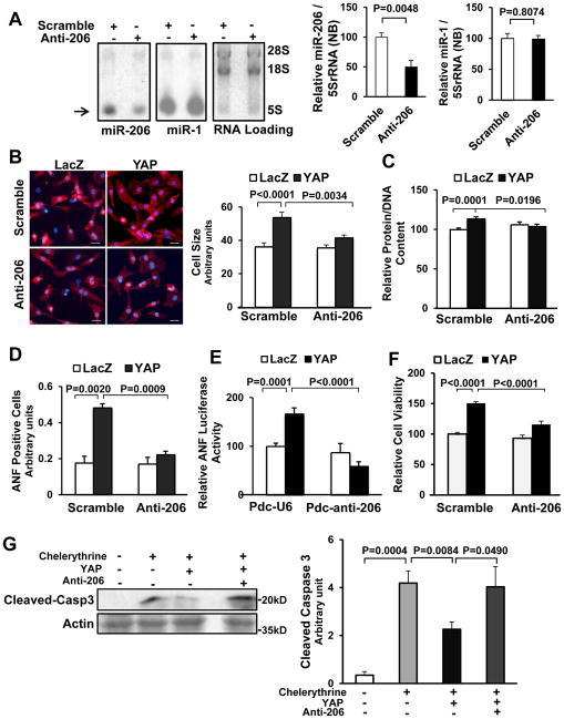
Figure 4. miR-206 mediates YAP-induced hypertrophy and inhibition of cell death.
Twelve to 24 hours after transduction of either Ad-sh-scr or Ad-anti-206, CMs were transduced with Ad-LacZ or Ad-YAP for 48 hours. (A) CMs were harvested for Northern blot, using a miR-206 probe or a miR-1 probe. RNA loading was evaluated with ethidium bromide staining. The level of miR-206 and miR-1 was normalized by that of 5S-rRNA. N=5. (B) CMs were stained with anti-α-actinin antibody (red) and DAPI (blue), and relative cell surface area (cell size) was evaluated. Scale bar, 30 μm. N=3. (C) CMs were harvested for the measurement of total protein content normalized by DNA content. N=3. (D) CMs were stained with anti-ANF antibody and DAPI. ANF-positive cells were quantified. N=3. (E) CMs were transfected with ANF luciferase reporter gene and either Pdc-U6 or Pdc-anti-206 plasmids before transduction with Ad-LacZ or Ad-YAP. After 48 hours, myocytes were harvested for ANF luciferase activity measurements. N=4. (F) CMs were harvested for Cell Titer Blue assays. N=3. (G) CMs were treated with or without chelerythrine (5 μM) for 1 hour and harvested for immunoblot analyses with anti-cleaved caspase 3 (Cleaved-Casp3) antibody and the results were quantified by densitometry. N=4.

