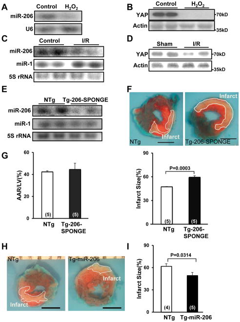
Figure 5. miR-206 protects the heart against I/R injury.
(A, B) CMs were treated with vehicle or H2O2 (50 μM) for 1 hour. Representative results of Northern blot analyses with miR-206 and U6 probes (A) or immunoblot analyses with anti-YAP antibody (B) are shown. N=4. (C, D) FVB mice at 3 months of age were subjected to 45 minutes of ischemia and 24 hours of reperfusion (I/R) or sham operation and the hearts were harvested for Northern blotting with miR-206 and miR-1 probes (C) and immunoblot analyses performed with anti-YAP antibody (D). N=5. (E) Tg-206-SPONGE and NTg mouse hearts were harvested for Northern blot analyses with miR-206 and miR-1 probes. N=3. (F, G) Tg-206-SPONGE and NTg mice at 3 months of age were subjected to 45 minutes of ischemia and 24 hours of reperfusion. The hearts were subjected to Alcian blue (1%) and TTC (1%) staining. In F, representative images of Alcian Blue dye and TTC staining are shown. Boundaries of the infarct areas are indicated by white lines. Scale bar, 2 mm. In G, area at risk (AAR)/LV (%) (left) and infarct size/AAR (%) (right) are shown. (H, I) Tg-miR-206 and NTg mice at 3 months of age were subjected to 45 minutes of ischemia and 24 hours of reperfusion. In H, representative images of Alcian Blue dye and TTC staining are shown. Boundaries of the infarct areas are indicated by white lines. In I, infarct size/AAR (%) is shown.

