Fig. 7.
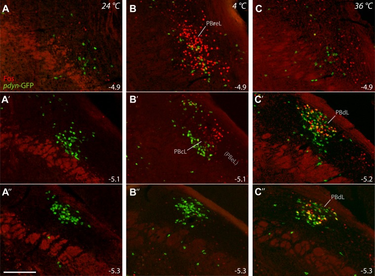
LPB neurons express c-Fos in distinct patterns after exposure to cool or warm ambient temperature. c-Fos immunofluorescence (red) and Pdyn-GFP fluorescence (green) are shown at three rostrocaudal levels of the LPB. A: control mice (24°C) have minimal c-Fos throughout the LPB. B: cool-exposed mice (4°C) express c-Fos in the far-rostral LPB, clustering in PBreL. Cool-activated, Fos+ neurons extend caudally into a rostral, lateral portion of the PBcL, as shown in B′, but are absent at caudal levels. C: Warm-exposed mice (36°C) express c-Fos primarily at caudal levels of the LPB. c-Fos labeling in warm-exposed mice colocalizes with and overlaps the dense cluster of Pdyn-GFP+ neurons in PBdL. Approximate bregma level (in mm) is indicated in each panel. Scale bar: 200 μm.

