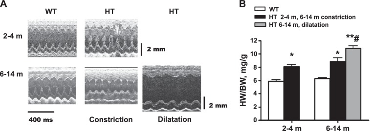
Fig. 2.
Left ventricular chamber remodeling in Marfan HT mice. A: representative sonographic (M-mode long-axis) images of the heart in Marfan HT mice and WT age-matched controls. Marfan HT mice display a mild cardiac hypertrophy at age 2–4 mo and cardiac hypertrophy with constricted (Con) or dilated (Dil) left ventricle at age 6–14 mo. B: quantification of ratio of heart weight (HW) to body weight (BW). Values are means ± SE; n = 6 (WT, 2–4 m), n = 7 (Marfan HT, 2–4 m), n = 13 (WT 6–14 m), n = 6 (Marfan HT, 6–14 m concentric hypertrophy), and n = 7 (Marfan HT, 6–14 m, dilatation). *P < 0.05 and **P < 0.01, WT vs. HT constriction. #P < 0.05, Dil vs. Con in 6- to 14-m age group.

