
Fig. 1.
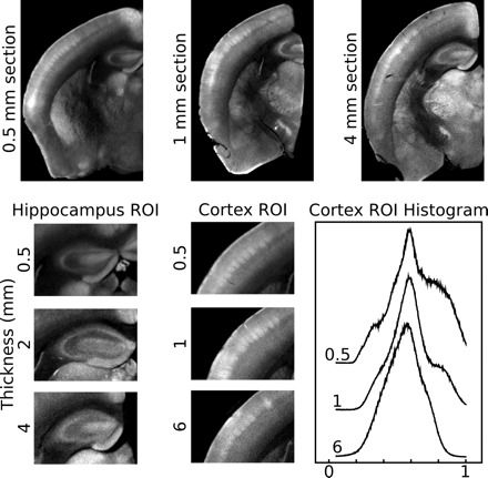
Effect of specimen size on image quality. Optical projection tomography (OPT) images of half-brains that were sectioned to be 0.5, 1, 4, or 6 mm thick are shown. With increasing sample thickness, the images are more blurred and the details are less resolved. This is highlighted in the both hippocampal and cortical regions of interest (ROI). In the hippocampal ROI there is a decrease in apparent image contrast with increasing thickness. This is also apparent in the cortical ROI, where the barrel field in layer 4 is less defined in the 6 mm sample compared with the 0.5 mm sample. This is further demonstrated in the cortical ROI histogram, where more at distinct intensities are observed in thinner samples.