
Fig. 4.
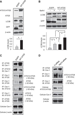
Competitive reactions between ATG5, ATG12, and Cav-1. A–C: Beas-2B cells were transfected with wild-type ATG5 or its control vector EGFP for 48 h (A and C), followed by BafA1 (100 nM) treatment for 2 h (B). Samples were then subjected to Western blot analysis (A and B) or IP assays (C) as indicated in the figures. Blots were quantified from 3 independent results. *Significant difference (P < 0.05). D: Beas-2B cells were transfected with ATG12-siRNA or its control for 48 h and subjected to IP assays as indicated. Cellular Cav-1 and β-actin derived from same cell lysates of IP experiments served as input controls.