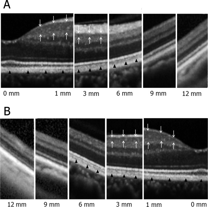
Fig. 3.

Highly magnified images of the normal retina from the macula to the periphery in nasal (A) and temporal (B) directions. In the macular region, there were six retinal layers and four bands in the outer retina that were distinguishable. Each layer became thinner as the scans became increasingly peripheral. In the peripheral retina as well as in the macula, most layers but the ganglion cell layer were visible. The ganglion cell layer (white arrows) was delineated in the macular region at 3 mm from the foveola but became undistinguishable from nerve fiber layer and inner plexiform layer in regions further than 3 mm eccentric from the fovea. The ELM, the second band, and the RPE were continuously delineated from the macula to the periphery, but the third band (black arrowheads) became indiscernible in the periphery.
