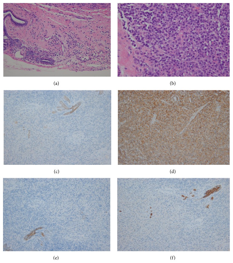
Figure 2.
Histopathological analysis ((a) HE ×200) of left breast mass shows infiltration of plasmacytoid cells around the ductal breast tissue. The plasmacytoid cells are eccentrically located with slightly enlarged nuclei compared to mature plasma cells ((b) HE ×400). Photomicrographs show a positive reaction for CD 138 ((c) ×200), indicating plasma cell origin, and positive reactions for E-cadherin ((d) ×200), CK5/6 ((e) ×200), and CK7 ((f) ×200) in the entrapped breast ductal tissue, but negative in infiltrated cells, suggesting a nonepithelial origin.

