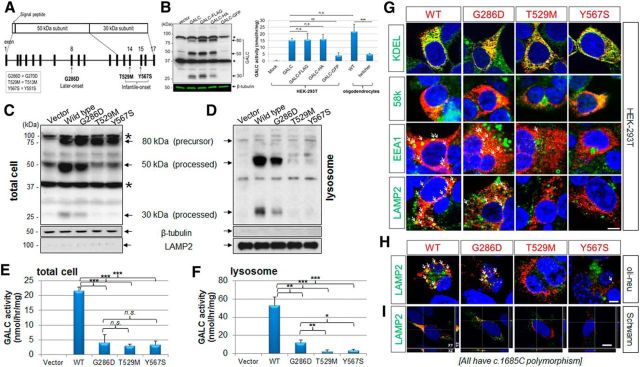
Figure 1.
GALC trafficking and processing in transfected cells is impaired in infantile-onset mutants but not in a later-onset mutant. A, A scheme is shown of GALC structure and common missense mutations found in GLD of European ancestry (Wenger, 2011a). The two most common infantile-onset missense mutations of GLD are T529M (exon 14) and Y567S (exon 15). The most frequent later-onset mutation is G286D (exon 8). B, Forty-eight hours after transfection of HEK-293T cells, total cell lysates were analyzed with a polyclonal anti-GALC antibody (Lee et al., 2010) that detects the 80 kDa precursor, and both the 50 and 30 kDa processed forms of GALC protein. There were nonspecific bands ∼90 and 40 kDa (asterisks). β-Tubulin was used for loading control. FLAG and HA fused to the C terminus of GALC were comparable with the untagged protein, in contrast fusion with eGFP markedly decreased GALC expression. GALC-FLAG and GALC-HA had similar enzyme activities to the nontagged GALC, whereas GALC-GFP had lower enzymatic activity. Oligodendrocyte GALC enzymatic activities at 4 DIV from WT and Galc-deficient twitcher mice, an authentic mouse model of GLD, were used for comparison. C, Compared with wild-type, the later-onset mutation (G286D) is significantly processed, whereas the infantile forms (T529M and Y567S) are much less processed. D, Lysosomal fractions, which were purified from the lysate of GALC-transfected HEK-293T cells and analyzed by Western blotting with anti-GALC, β-tubulin, and LAMP2 antibodies, contain mostly the processed forms of GALC, but not the precursor form. In the lysosomal fractions, there are almost no processed forms of GALC for the infantile forms (T529M and Y567S) in contrast to the later-onset form (G286D). E, F, In total cell lysates, the GALC activity of the later-onset mutant (G286D) is indistinguishable from that of the infantile forms (T529M and Y567S), whereas in lysosomal fractions, there is residual GALC activity for the later-onset mutant, but almost no activity for the infantile-forms, revealing a correlation between lysosomal GALC activity and severity of disease. Error bars indicate SD of triplicates. *p < 0.05 (Student's t test). **p < 0.01 (Student's t test). ***p < 0.001 (Student's t test). n.s., Not significant. G, Immunofluorescence for WT or the three mutant GALCs in HEK-293T cells with anti-GALC (red) and anti-KDEL, anti-58k, anti-EEA1, or anti-LAMP2, antibodies (all green) showed that WT and later-onset mutant GALC colocalized with all four subcellular markers, whereas the infantile-onset GALC mutants were not (or minimally) detectable in the lysosome, and mainly found in the ER and Golgi. The same analyses in transfected oli-neu cells at 4 DIV (H) and primary rat Schwann cells (I) with anti-GALC (red) and anti-LAMP2 (green) confirmed that the WT GALC and later-onset mutant are present in the lysosome, whereas the infantile-forms are not. Arrows indicate overlapping lysosomal puncta and GALC (yellow). Orthogonal views (XY, XZ, and YZ) of a z-stack are shown. White lines indicate section positions. Nuclei were labeled with DAPI. DIV, Days of in vitro differentiation. Scale bars: G–I, 5 μm.

