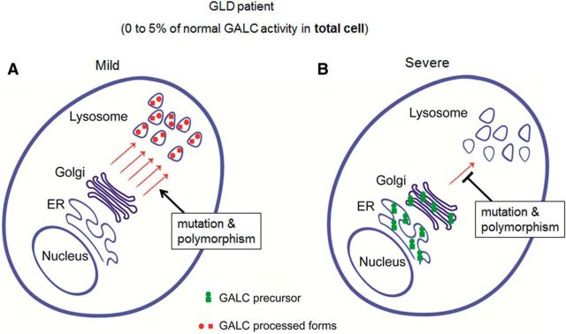
Figure 9.
Model for how measured GALC activities may not correlate with disease severity in GLD; 5% of normal GALC activity is regarded as the general cutoff for diagnosis of GLD. The diagram represents two different possibilities for the localization of GALC that can produce the same whole-cell lysate GALC activity when measured in patient blood. A, If the majority of remaining GALC is <5% but is found concentrated in the lysosome, then the patient will have milder phenotype and present with a later-onset form of the disease. B, If, however, most of the remaining GALC is localized to the ER or Golgi, due to defective trafficking, then the patient will have a more severe phenotype and may manifest with the infantile-onset form of the disease.

