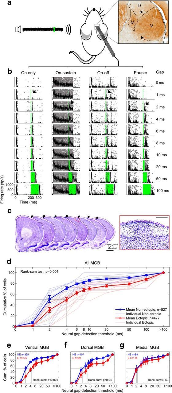
Figure 1.

Ectopic mice have a deficit in auditory thalamic sensitivity to brief gaps in noise. a, Extracellular in vivo recordings were obtained from neurons in the ventral (V), dorsal (D), and medial (M) subdivisions of the MGB of anesthetized mice during presentations of gap-in-noise stimuli with variable gap durations. Inset, Coronal section through MGB, stained for cytochome oxidase. Arrowheads indicate electrolytic lesions. b, Examples of four common types of auditory thalamic responses to gap-in-noise stimuli, shown in rasters with superimposed PSTHs. Zero time corresponds to start of noise; firing rate is shown in spikes per second (sp/s). Green column represents silent gap. Black arrow indicates neural gap-detection threshold. c, Example neocortical ectopia in motor cortex, shown in successive coronal sections (left, arrows; scale bar, 1 mm) and in a magnified view of layer I (right; scale bar, 200 μm). d, Neural gap-detection deficit in MGB of ectopic mice. Red represents ectopic mice. Blue represents nonectopic mice. Dotted lines indicate individual animals. Solid lines with error bars indicate mean ± SE across animals. Dotted gray lines indicate median values. n, total number of neural recordings across animals. e–g, Same analysis for neurons recorded in ventral MGB (e), dorsal MGB (f), and medial MGB (g). Results of Wilcoxon rank-sum tests for differences in medians are shown. N.S., Not significant. Kolmogorov–Smirnov tests for differences in distributions produced similar results (see Results).
