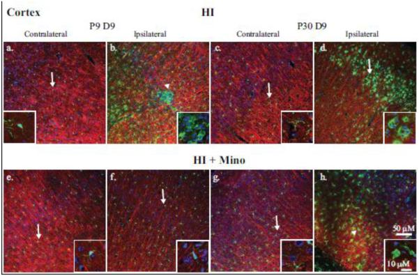
Figure 4. Immunohistological analysis of microglial response in the cortex.
Double immunohistological staining for Iba1 (green) and MAP2 (red) proteins were examined in the cortex. Nuclei were stained with DAPI (blue). Upper panels: the contralateral (a, c) and ipsilateral (b, d) cortex of P9 (a, b) and P30 (c, d) brains at 9 days post-HI are compared. Lower panels: the contralateral (e, g) and ipsilateral (f, h) cortex of P9 (e, f) and P30 (g, h) brains at 9 days post-HI from minocycline-treated mice are compared. Insets: magnification (240X) of representative microglia demonstrating either ramified (arrow) or amoeboid (arrowhead) morphology.

