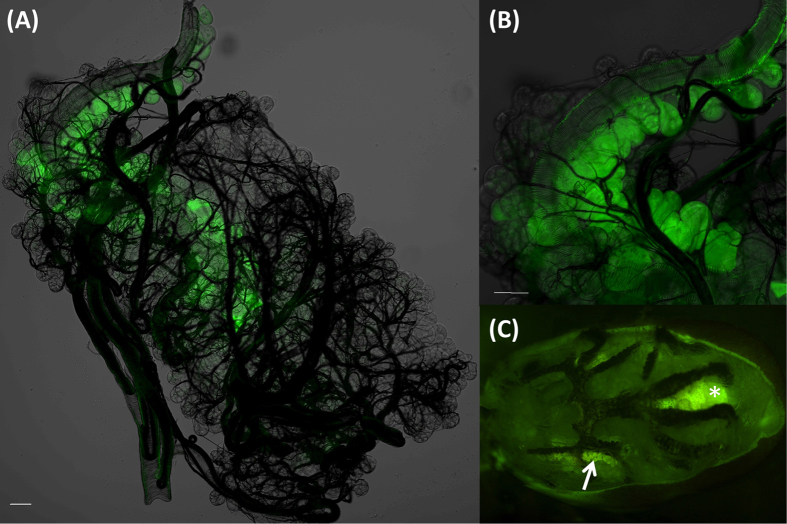
Figure 8. Absorptive function of type I acini shown by absorption of Rhodamine 123.
Ticks were dissected to trace the fluorescence after the desiccated tick was offered a drop of water containing Rhodamine 123 (1 mmol l−1) for ~5 hr, (A) Intact salivary glands of unfed tick. (B) Magnified view of type I acini. (C) Overview of fluorescence from whole unfed tick body after the tergum was removed. Arrow in (C) indicates the salivary glands. Asterisk indicates hindgut and rectal sac that is autofluorescent (See Supplementary Fig. S6). Scale bars equal 50 μm.

