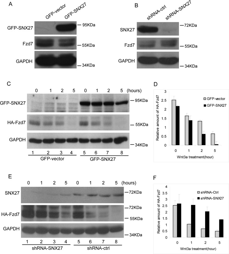
Figure 4. SNX27 promotes the degradation of Fzd7.
(A) MCF7 cells stably expressing GFP or GFP-SNX27 were lysed and processed for western blot to detect Fzd7 using polyclonal antibody against Fzd7, showing less amount of Fzd7 in cells expressing GFP-SNX27 compared with that of cells expressing GFP (middle panel). (B) MCF7 cells stably expressing shRNA-ctrl or shRNA-SNX27 were lysed and processed for western blot to detect Fzd7 using polyclonal antibody against Fzd7, showing that Fzd7 is efficiently depleted (upper panel) and the amount of Fzd7 in SNX27 knockdown cells is much more than that of control cells (middle panel). (C) MCF7 cells were co-transfected with HA-Fzd7 and GFP vector or GFP-SNX27, then induced for degradation with Wnt3a for the indicated time, in the presence of cycloheximide. The cell lysates were subjected for western blot to detect HA-Fzd7 using HA-tag antibodies. The results demonstrated that over-expression of SNX27 promotes the degradation of Fzd7 (meddle panel). (D) Quantitative analysis of the results from three independent experiments represented in C showing that over-expression of SNX27 promotes the degradation of Fzd7. (E) MCF7 cells expressing shRNA-SNX27 were transfected with HA-Fzd7 then induced for degradation with Wnt3a for the indicated time. HA-Fzd7 was detected by similar approach mentioned above. The results demonstrated SNX27 knockdown significantly inhibits the degradation of Fzd7 (meddle panel). (F) Quantitative analysis of the results from three independent experiments represented in E showing that SNX27 knockdown inhibits the degradation of Fzd7.

