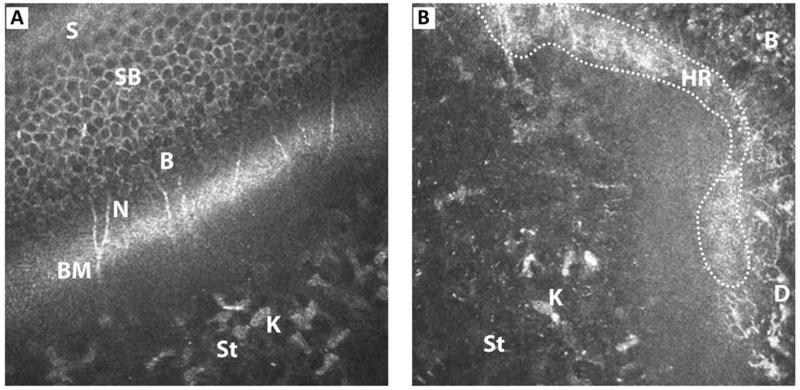
FIGURE 4. Oblique section of (A) corneal epithelium and (B) limbal epithelium through their anterior stroma, showing all layers.
B - basal epithelium; BM - Bowman’s layer; D - dendritic cells; K - keratocytes; N - nerve plexus; HR - hyperreflective region; S - superficial epithelium; SB - suprabasal epithelium; St - anterior stroma.

