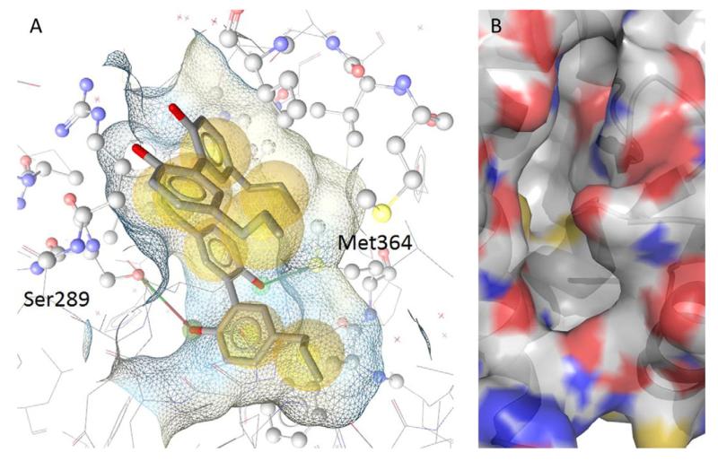
Fig. 2.
(A) Two molecules of magnolol concomitantly occupying the binding site of PPARγ (PDB 3R5N) are shown, with the chemical interaction pattern that defines the activity of the molecules depicted. Yellow spheres represent hydrophobic interactions, red and green arrows mark hydrogen bond acceptor and donor atoms. This interaction pattern may be converted into a structure-based pharmacophore model and used for virtual screening. (B) The empty binding pocket of PPARγ is shown, which can be used in docking simulations to place new molecules into the binding site and to calculate the binding free energy of the ligand.

