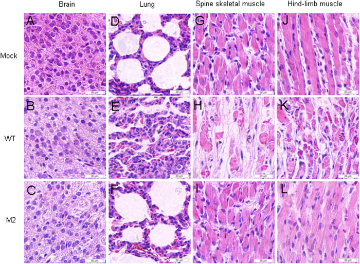
Figure 5. Pathological analysis of WT or M2 infected neonatal mice.
One-day-old ICR mice were intracerebrally inoculated with medium or WT or M2 virus from transfected HEK293T cells at 104 CCID50 ml−1. No histological change was observed in the brain of the non-infected medium control (A) or WT (B) or M2 (C) infected mice. No histological change was observed in the lung (D,F) or hind limb muscle (G,I) or spinal muscle (J,L) of the non-infected medium control or M2 infected mice. Mice infected with WT viruses exhibited severe alveolar shrinkage (E) in the lung tissue. Mice infected with WT viruses (grades 4 to 5) exhibited signs of severe necrosis, including muscle bundle fracture, dissolution of muscle fiber cells, nuclei shrinkage and swelling (H and K). A to L, magnification 400× .

