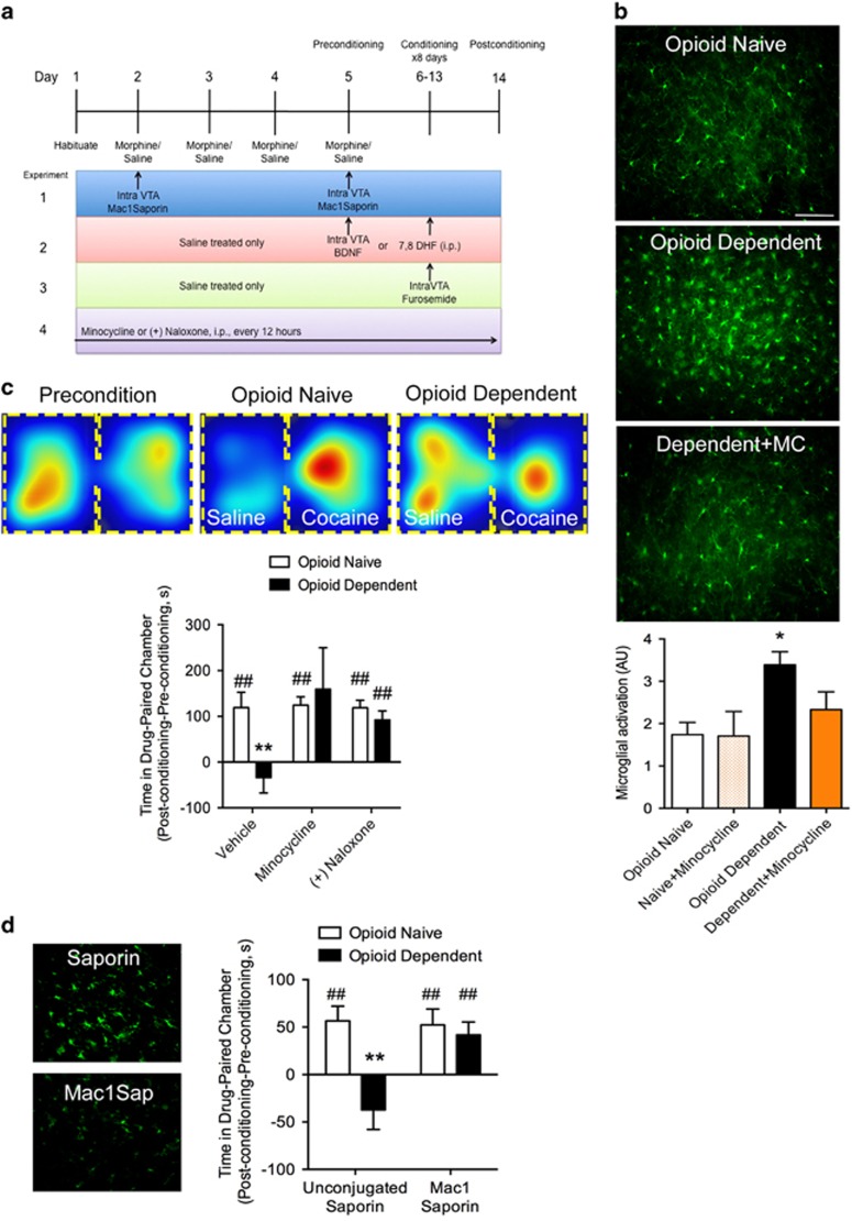
Figure 1.
Chronic opioid treatment activates microglia and interferes with cocaine place preference. (a) Experimental design. (b) Microglia, identified by an antibody against IBA-1, were labeled in the VTA. Microglial activation was quantified based on defined morphological criteria, including cell body size and ramified branching. A chronic, ascending dose regimen of morphine resulted in significant microglial activation that could be blocked by daily systemic administration of the microglial inhibitor, minocycline (MC). Scale bar =100 uM. Bar graph represents quantification of activated microglia in VTA sections. n=4–5. Error bars=SEM, *p<0.05, n=3–12. (c) Top images display a heat map of total time spent in either the drug-paired or saline-paired chamber before (precondition) and after training. Bottom histograms plot the difference in time spent on the drug-paired side on the pre-conditioning day and the post-conditioning day. Opioid-dependent animals did not show a place preference to the cocaine-paired chamber. Systemic minocycline or (+)-naloxone, the opioid-inactive enantiomer previously shown to inhibit microglial activation, treatment recovered a cocaine place preference in opioid-dependent animals, but had no effect on cocaine place preference in opioid-naïve animals. Error bars=SEM, **p<0.01 when compared with opioid-naïve, ##p<0.01 when compared with baseline, n=5–10. (d) Animals in which VTA microglia were depleted with Mac-1-Saporin retained cocaine place preference in opioid-dependent animals. Mac-1-Saporin did not affect the development of cocaine place preference in naïve animals. Injection of unconjugated saporin had no effect on the development of cocaine place preference in naïve animals and it did not restore cocaine place preference in opioid-dependent animals. Error bars=SEM, **p<0.01 when compared with opioid-naïve, ##p<0.01 when compared with baseline, n=8–10. Photomicrographs depict IBA-1 labeling in VTA sections from animals treated with unconjugated saporin or Mac-1-Saporin. Scale bar=100 uM.

