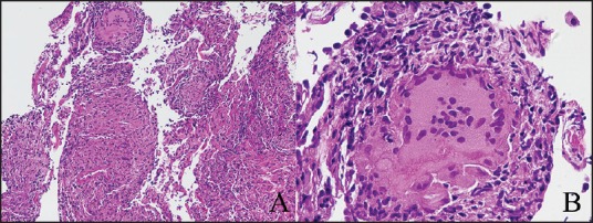
Figure 3.

Transbronchial biopsy (TBB) showing granulomas. H and E, assay ×100 (a), ×200 (b). TBB was mainly characterized by the presence of nonnecrotizing granulomas. Other inflammatory cells were poorly represented, mainly lymphocytes around granulomas. Alveolar septa were overshadowed by granulomatous component. Some central necrosis (b) revealing very active sarcoidosis can be identified. Special stains for fungi (PAS) and mycobacteria resulted negative. Images courtesy of Tironi Andrea MD, Division of Anatomic Pathology, Spedali Civili, Brescia.
