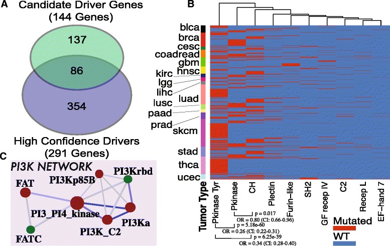
Fig. 2.

a Venn diagram of the represented Pfam domains in the list of 291 high confidence drivers and 144 candidate drivers. A total of 577 different Pfam domains are covered by these genes with 86 Pfam domains shared between the two lists. b Heatmap representation of significant Pfam domains in the “Kinase” network. Every row represents a patient of 17 different tumor types. A strong mutual exclusivity between tyrosine kinases, kinases and CH domain is shown. c PI3K networks in driver genes. Every circle represents a distinct Pfam domain and the size represents the number of genes that contain the specified Pfam domain. Color indicates if significant hotspots were found in the LowMACA analysis (red is significant, green is not significant). Two domains are connected if they are found together on the same gene/protein. Edge thickness represents the number of genes that harbor both Pfam domains at the vertices (minimum 2). Blue color indicates mutual exclusivity and orange depicts significant co-occurrence
