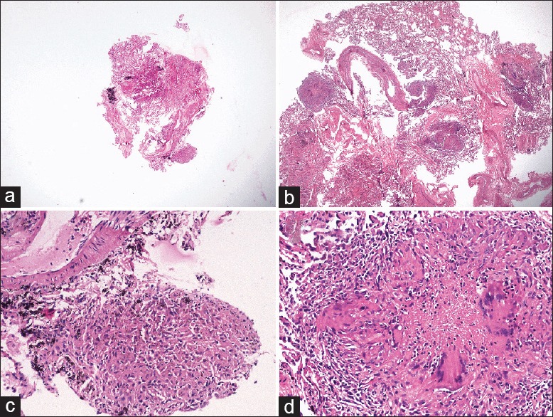
Figure 3.

(a and b) Photomicrographs showing comparison of sizes of the flexible FB and CB under scanner view [hematoxylin and eosin (H and E), ×40], (c) FB showing single focus of loose collection of epithelioid cells forming granuloma (H and E, ×200), (d) CB showing well-formed epithelioid cell granuloma with Langhans giant cells and central necrosis with nuclear debris (H and E, ×200)
