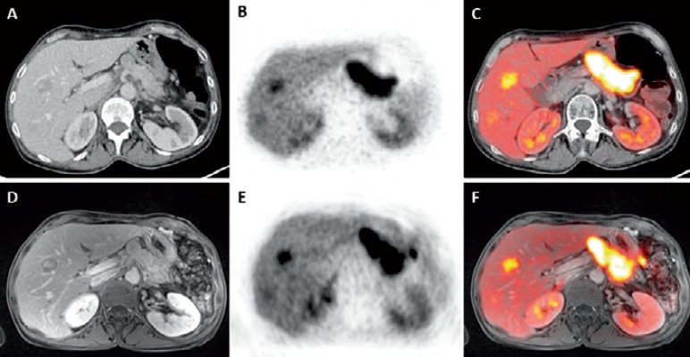
Fig. 8.
A 62-year-old female patient with a NET liver metastasis in the right liver lobe on CT. B This liver lesion shows increased (68Ga)-DOTATOC signal on PET and C on the fused DOTATOC-PET/CT. D The same liver metastasis of the same patient on contrast-enhanced T1w MR image, E clearly visible on PET, and F on fused (68Ga)-DOTATOC-PET/MRI.

