
Figure 3. Recombinant perlecan Dm IV cleavage by MMP-7.
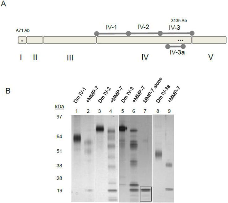
A) Diagram of recombinant subdomains Dm IV-1, 2, 3, and Dm 3a compared to full length perlecan. B.) Digestion of recombinant domain IV protein by MMP-7. Various subdomains of Dm IV: IV-1 (lanes 1,2), Dm IV-2 (lanes 3, 4), Dm IV-3 (lanes 5, 6), Dm IV-3a (lanes 8,9) were incubated alone or with MMP-7 (1 μg of Dm IV with 0.1 μg MMP-7) overnight at 37°C. Shown is a silver stain after SDS-PAGE separation on a 4–12% (w/v) acrylamide gradient gel. MMP-7 can cleave every subdomain. The black boxed portion in lane 7 (MMP-7 alone) indicates the MMP-7 band.