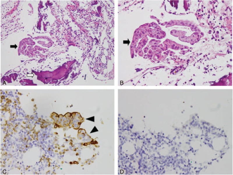
FIGURE 5.

Histologic findings of metastatic adenocarcinoma at L2 vertebral body. (A) and (B) Hematoxylin and eosin stain (A: 10 × 20, B: 10 × 40): metastatic adenocarcinoma, composed of tumor cells (black arrow) with large, pleomorphic nuclei, prominent nucleoli, arranged in tubular or small glandular structure. Bone (white arrow); (C) immunohistochemical stain shows positive to carcinoembryonic antigen (arrow head) (10 × 40); (D) immunohistochemical stain shows negative to thyroid transcription factor-1 (10 × 40).
