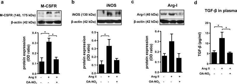
Figure 5. OA-NO2 decreases the Ang II-induced fibrotic processes in heart tissue.
The expression of M-CSFR (a), iNOS (b), and arginase-I (c) was detected in heart tissue of C57BL/6J mice treated for 2 weeks with Ang II (1.5 ng/g/min) and OA-NO2 (6 mg/kg) via subcutaneously implanted osmotic minipumps. The concentration of TGF-β was determined in plasma samples (n=6-9) (d). The pictures represent one of several individual experiments (n=6-9). A *p value of less than 0.05 was considered significant when evaluating differences between the individual bars and positive control (Ang II-treated mice).

