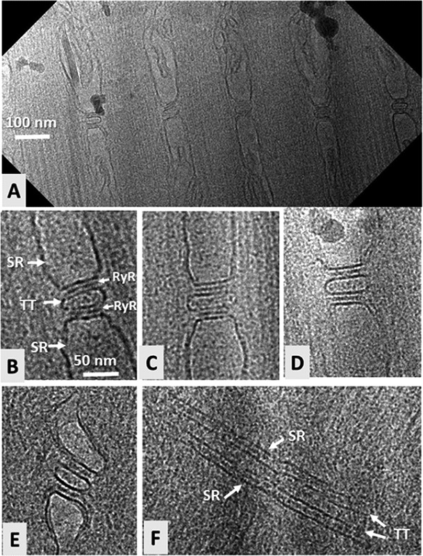
Fig 2.

Triad junctions imaged by cryo-electron microscopy of FIB-milled muscle. A. Overview of a 300-nm-thick longitudinal section showing five triad junctions from toadfish swimbladder. B-D. Higher- magnification images of three triad junctions. Abbreviations: SR, sarcoplasmic reticulum; TT, T-tubule; RyR, ryanodine receptor. E-F. Triad junctions from zebrafish skeletal muscle. E shows a longitudinal view with the T-tubule in cross-section (as in A-D), while F shows a longitudinal view in which the T-tubule is viewed from the side (arrows adjacent to “TT” indicate the two opposing surfaces of the membrane of the T-tubule); rows of RyRs are visible in the two spaces between the TT and SR membranes. A and B adapted from reference.13
