Fig 1.

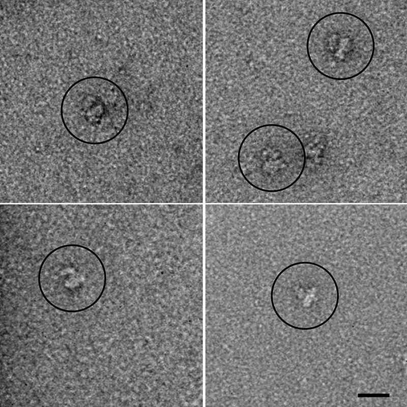
Electron microscopy of negatively stained DHPR. The main globular body with an appendage is visible in the raw images. Scale bar, 20 nm.

Electron microscopy of negatively stained DHPR. The main globular body with an appendage is visible in the raw images. Scale bar, 20 nm.