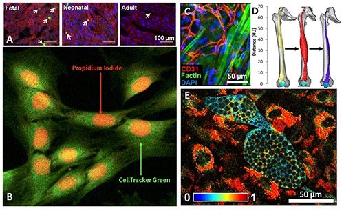
Fig 1.

Types of functional assessment metrics for various tissue engineering studies. A) Proliferation of cardiomyocytes measured by immunohistochemical staining of phosphohistone H3 15. B) Muscle cells viability measured in cells stained with CellTracker™ Green and propidium iodide showing dying myocytes.136 C) Periphery of implanted engineered muscle showing neonatal rat satellite cells and their myogenic predifferentiation via f-actin filamentous formation and integration with extant, CD31-labeled endotheilial cells.137 D) CT reconstructions of denervated thigh muscle that was electrically stimulated for growth, highlighting a possible modality for measuring myogenesis in engineered muscle tissue.143,144 E) Changes in redox ratios observed in adipogenic differentiation with TPEF along with characteristic, non-autofluorescent lipid droplets.40
