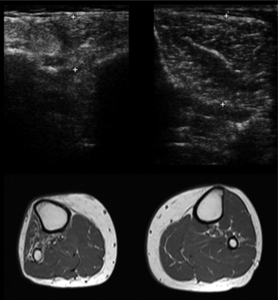
Fig 2.

Fatty replacement of the anterolateral compartment muscles of the leg following common fibular nerve injury: ultrasound and magnetic resonance axial images at the level of the middle third of the tibialis anterior belly. The right anterolateral compartment muscles are denervated, so, compared to the contralateral ones, these muscles are atrophic with increase of intramuscular fat: in the ultrasound images, there is a marked thinning of the tibialis anterior muscle with increased echogenicity and loss of physiological heterogeneity of the ecostructure; in the T1-weighted MR image, these muscles are brighter.
