
Fig 3.
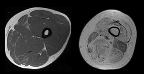
Fatty degeneration: axial T1-weighted images of the thigh showing, on the left, the normal muscular appearance on MR, characterized by a homogeneous grayish ("isointense") signal intensity. On the right, there is a severe and diffuse muscular fatty degeneration: these muscles are hyperintense compared to the previous ones because of the increase of intramuscular fat.