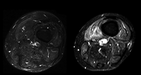
Fig 4.

Intramuscular edema: axial STIR images of the thigh showing, on the left, normal muscle signal intensity. In this sequence, the signal coming from the fat is suppressed, so the fat-containing structures are hypointense; on the right, the muscles are hyperintense, particularly the vastus medialis muscle due to the increase of water typical of inflammatory changes.
