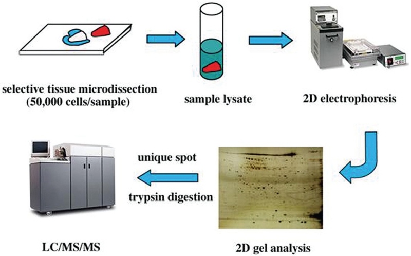
FIGURE 1.
Drawings and photographs showing the steps involved in tissue microdissection and proteomic profiling. Selective tissue microdissection ensures the purity of the tissue sample being analyzed. Once this is completed, the sample is broken (or lysed) into its individual protein components. When placed on a 2-dimensional (2D) gel with an electric current, the proteins will settle at distinct locations based on their charges and molecular weights. These locations are used to generate a “map” of the protein composition on the 2D gel. Further characterization is completed using liquid chromatography (LC), mass spectroscopy (MS), or a combination of the 2 methods.

