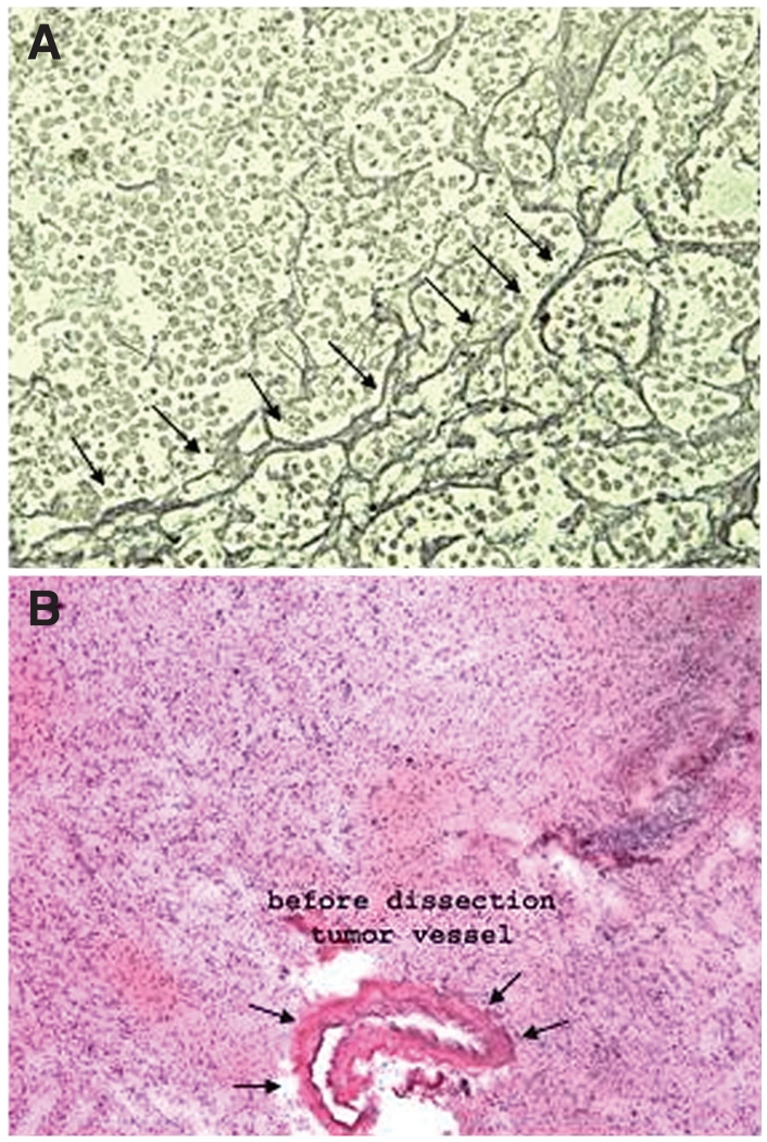
FIGURE 2.
Photomicrographs illustrating that neurological disorders often have distinct boundaries that differentiate them from normal nervous tissue. A, the reticulin border of a pituitary adenoma (arrows) can be used as a capsule in isolating the adenoma for proteomic analysis, while excluding the normal pituitary gland. B, a blood vessel surrounding a high-grade glioma can be used to analyze the unique proteins associated with glioma vasculature. This can then be used to characterize proteins that are unique to a particular glioma subtype or grade.

