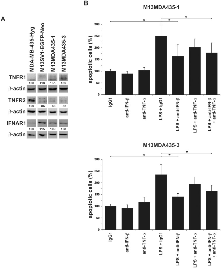
Fig 5. Neutralization of IFN-β, but not TNF-α, impaired the LPS induced apoptosis in M13MDA435-1 and -3 hybrid cells.
A) Western Blot analysis revealed comparable TNFR1 and IFNAR1 expression levels in the investigated cell lines. By contrast, TNFR2 expression levels were markedly lower in M13SV1-EGFP-Neo human breast epithelial cells and M13MDA435-1 and -3 hybrid cells. Shown are representative data of three independent experiments. The relative protein expression levels were calculated in relation to the appropriate β-actin expression level, whereby MDA-MB-435-Hyg cells were set to 100%. B) M13MDA435-1 and -3 hybrid cells were cultivated in the presence of LPS (100ng/ml) and neutralizing IFN-β and TNF-α antibodies (10μg/ml) for 24h. The relative amount of apoptotic cells was calculated in relation to the IgG1 control, which was set to 100%. Shown are the mean ± S.E.M. of five independent experiments. Significance: * = p<0.05.

