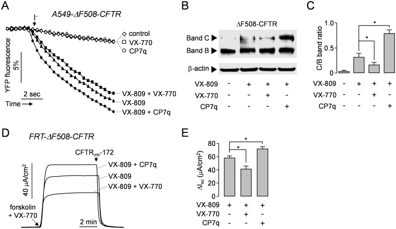
Fig 6. CP7q increases the functional rescue of ΔF508-CFTR by VX-809 in A549 and FRT cells.
(A) ΔF508-CFTR expressing A549 cells were incubated with 5 μM VX-809 in the presence and absence of 10 μM CP7q or 5 μM VX-770 for 48 hours. The traces are showing iodide influx via the rescued ΔF508-CFTR stimulated with 10 μM forskolin and 10 μM VX-770 (mean ± S.E., n = 6). (B) Representative ΔF508-CFTR immunoblot at 48 h after treatment with 5 μM VX-809 in the presence and absence of 10 μM CP7q or 5 μM VX-770 in A549-ΔF508-CFTR cells. (C) The ratio of ΔF508-CFTR band C/B was summarized in bar graph (mean ± S.E., n = 3). (D) Representative apical membrane current traces showing effect of CP7q and VX-770 on VX-809-induced functional rescue of ΔF508-CFTR in FRT-ΔF508-CFTR cells. Well differentiated FRT cells were incubated with 5 μM VX-809 in the presence and absence of 10 μM CP7q or 5 μM VX-770 for 48 hours. ΔF508-CFTR currents were inhibited by 10 μM CFTRinh-172. (E) Bar graph showing the summarized data of peak current (mean ± S.E., n = 6). *P < 0.05.

