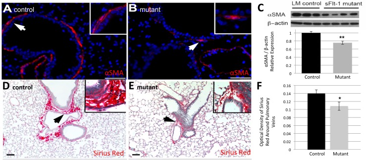
Fig 4. Reduced VEGF disrupts the parenchyma around airways and vasculature.
(A, B) Representative images of immunofluorescent staining for α smooth muscle actin (αSMA) reveals fewer putative myofibroblasts surrounding the airways in mutant mice (B) compared to controls (A). (C) Quantification by Western blot confirms a statistically significant decrease in the relative abundance of αSMA in mutants. (D, E) Representative images of Sirius red stain for Type 1 collagen demonstrates decreased peri-vascular staining in mutants (E) compared to controls (D). (F) Mutant mice express significantly less collagen around pulmonary veins based on optical density quantification. White and black arrows point to magnified areas shown in insets. Scale bars represent 50 μm. Data are expressed as mean ± SD, *P< 0.05, **P< 0.01 versus control.

