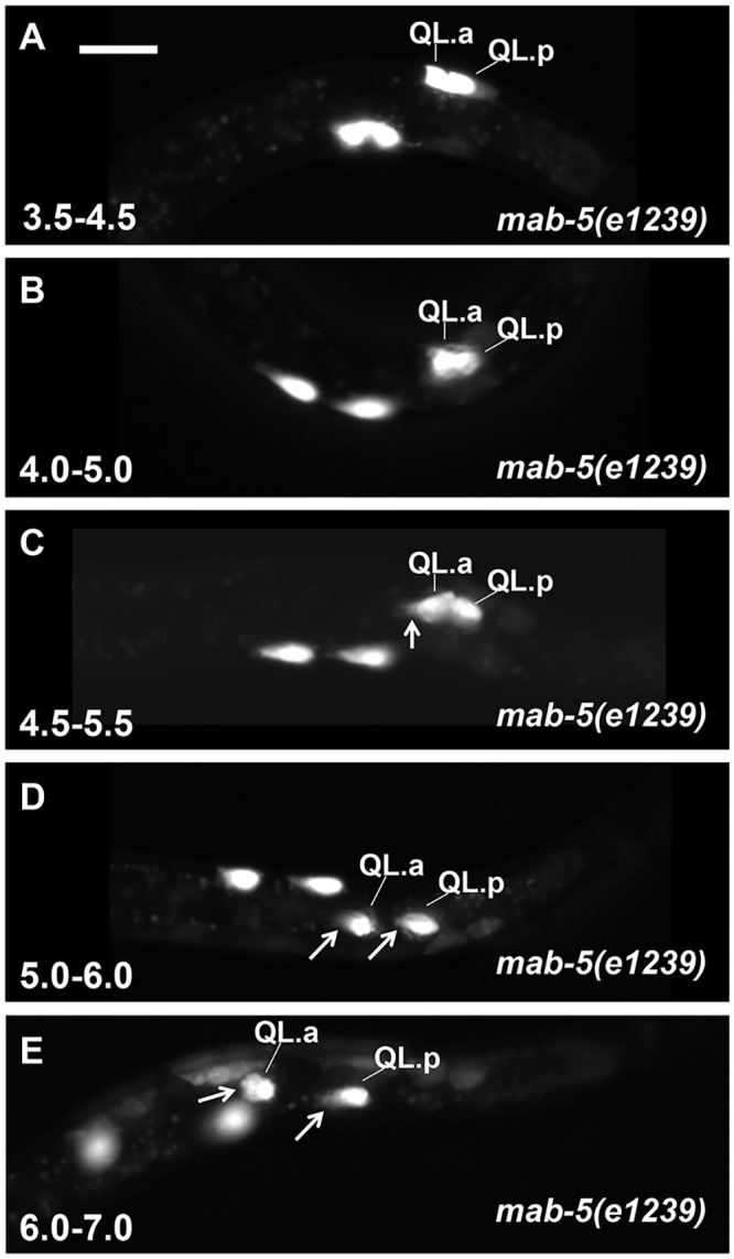
Fig 7. Q descendant migrations in mab-5 loss-of-function.

Fluorescent micrographs of ayIs9[Pegl-17::gfp] expression in the Q cells of mab-5(e1239) loss-of-function mutants are shown. At 4.0–5.0h and 4.5–5.5h, QL.a/p have not migrated, but QL.a is beginning to extend an anterior protrusion (arrow in C). Beginning at the 5.5–6.5h timepoint, both QL.a and QR.a begin posterior migration. Anterior is to the left, and the scale bar in A represents 5μm.
