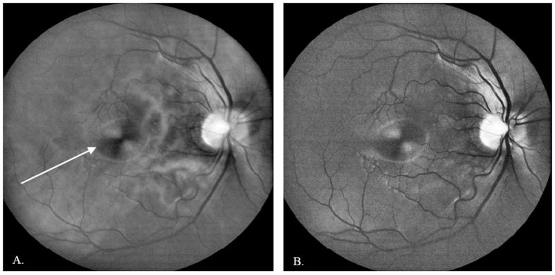
Figure 9.

50-frame averaged DLO image of an undilated 28 year old normal Asian male imaged at the Indiana University School of Optometry. A: Red channel image, acquired with red illumination (630 nm). White arrow shows the macular bow tie pattern caused by the birefringence in Henle’s fiber layer. B: Green channel image, acquired with yellow illumination (580 nm). The red illumination penetrates the deeper retina, providing visualization of the choroidal vasculature, whereas the yellow illumination has higher blood vessel contrast and provides better visualization of the superficial retina and nerve fiber layer.
