Figure 1.
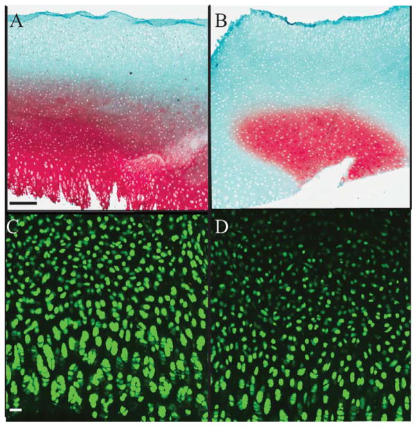
Trypsin treatment sufficiently removes native aggrecan from bovine ex vivo tissue. Safranin O & Fast Green staining (red) for GAG in bovine explants indicates <50% of residual GAG remains in aggrecan depleted matrices (Figure 1B) as compared to normal cartilage (Figure 1A). Live (green)-dead (red) stain of chondrocytes in the explant demonstrate living cells (green) in both explants indicating no death post trypsin treatment (Figure 1C, D). Scale bar for Figure A&B: 200μm, Figure C&D: 50 μm

