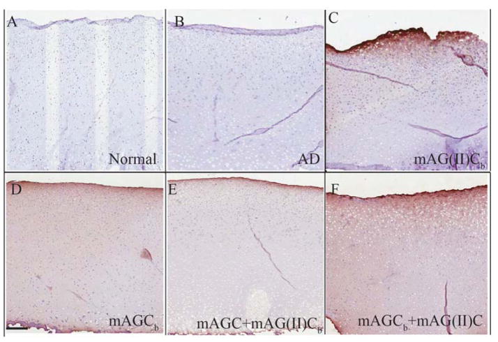
Figure 2.
Diffusion of molecules through bovine cartilage explant. Streptavidin HRP counter stained with diaminobenzidine was used on all experimental groups to probe for biotin labeled mAG(II)Cb (collagen type II binding) (figure 2C), mAGCb (HA binding) (figure 2D), mAGC + mAG(II)Cb (figure 2E) and mAGCb + mAG(II)C (figure 2F) diffused through aggrecan depleted cartilage explants. Normal cartilage (figure 2A) and aggrecan depleted cartilage (figure 2B) were treated with 1X PBS. Images represent the mid sagittal section of the explant and represent diffusion from articular surface towards the deeper zone. Scale bar 200 μm

